Reducing animal experiments: 3 R's grant for Guy Roukens

logo_ivd_en.gif

The process of cancer metastasis is a complex one and involves multiple steps. It involves cancer cells leaving the primary tumor and moving into the blood vascular system (intravasation), traveling to a distant site in the body, often the lungs, bone or brain. Here the tumor cells need to move out of the blood vessels  (extravasation) and then settle and grow into a secondary tumor (metastasis). Until now it has been very difficult to study these aspects of cancer biology in the lab, and almost all studies tend to use animal models. However, with recent developments in tumor-organoid culture and vascularization, together with microfluidic technologies, the possibility to develop in vitro systems for studying metastasis is becoming a possibility. 

At Utrecht University, the Animal Welfare Body awards research grants for projects aim at reducing the use of animal experiments. Guy Roukens, senior postdoc in the Coffer Lab, has been awarded such funding to explore the possibility of developing in vitro microfluidic systems that can be used to investigate tumor vascularization, intravasation and extravasation and thereby help understand the process of tumor metastasis.

Mesenchymal stem cell-derived extracellular vesicles can promote cartilage regeneration

Vonk et al. 20746n1_1-1.jpg

Osteoarthritis (OA) is a rheumatic disease leading to chronic pain and disability with no effective treatment available. Recently, allogeneic human mesenchymal stem cells (MSC) entered clinical trials as a novel therapy for OA. Increasing evidence suggests that therapeutic efficacy of MSC depends on paracrine signaling. Here we investigated the role of extracellular vesicles (EVs) secreted by human bone marrow-derived MSC (BMMSC) in human OA cartilage repair. We show that BMMSC-EVs inhibit the adverse effects of inflammatory mediators on cartilage homeostasis. BMMSC-EVs also promoted cartilage regeneration in vitro. Addition of BMMSC-EVs to cultures of chondrocytes isolated from OA patients stimulated production of proteoglycans and type II collagen by these cells. These data demonstrate that BMMSC-EVs can be important mediators of cartilage repair and hold great promise as a novel therapeutic for cartilage regeneration and osteoarthritis.

This work by Magdalena Lorenowicz is part of a combined research program with the Saris-Vonk Lab and has been accepted for publication in Thernostics.

Foxp1 regulates neurogenesis by modulating the Notch pathway

Foxp1_NSC.jpg

Mutations in Foxp1 have been linked to neurodevelopmental disorders including intellectual disability and autism, however, the underlying molecular mechanisms remain ill-defined. Here work from Luca Braciolli demonstrates utilizing RNA- and chromatin immunoprecipitation (ChIP)-sequencing that Foxp1 directly regulates genes controlling neurogenesis. We show that Foxp1 is expressed in embryonic neural stem cells (NSCs) and modulation of Foxp1 expression impacts both neuron and astrocyte differentiation. Using a murine model of cortical development, Foxp1-knockdown in utero was found to reduce NSC differentiation and migration during corticogenesis. Furthermore, transplantation of Foxp1-knockdown NSCs in neonatal mice after hypoxia-ischemia (HI) challenge demonstrated that Foxp1 is also required for neuronal differentiation and functionality in vivo. Foxp1 was found to repress the expression of Notch pathway genes including the Notch-ligand Jagged1, resulting in inhibition of Notch signaling.  Finally, blockade of Jagged1 in Foxp1-knockdown NSCs rescued neuronal differentiation in vitro. Together these data support a novel role for Foxp1 in regulating embryonic NSC differentiation by modulating Notch signaling.

This work is part of a collaboration with the Nijboer and Pasterkamp Labs and the full publication in Stem Cell Reports can be read here.


Congratulations Dr Ana Rita Lourenço: PhD survivor !

Ana-Rita Lourenco2.jpg

Transcriptional mechanisms regulating oncogenic TGF-β signaling in breast cancer

A complex program of epithelial-to-mesenchymal transition (EMT) plays a pivotal role during both embryogenesis and tissue homeostasis. However abnormal activation of this process can lead to tumor progression and metastasis. TGF-β signalling has been demonstrated to induce and support EMT by regulating a complex network of transcription factors. During this process, transcription factors responsible for maintaining the epithelial phenotype are supressed, whereas transcription factors involved in the acquisition of mesenchymal traits are rapidly induced. Such transcription factors are so called EMT-inducers (or EMT master regulators) and include family members of Zeb, Snail and Twis1t transcription factors.

Work in this thesis investigates the role of the transcription factor SOX4 as a crucial regulator of TGF-β-mediated induction of mesenchymal markers, and supports a tole for SOX4 as an EMT ‘master regulator’. Furthermore, the C/EBPα transcription factor was identified as a novel epithelial ‘gate-keeper’, inhibiting EMT. These two transcription factors can therefore control the balance between epithelial and mesenchymal transition in both organogenesis and development of disease, such as cancer metastasis.

Taken together our results provide novel insights into the transcriptional regulation of EMT and how this can be deregulated in disease. This identifies novel molecular pathways that could be therapeutically targeted to control epithelial-mesenchymal cell differentiation as well as tumor metastasis.

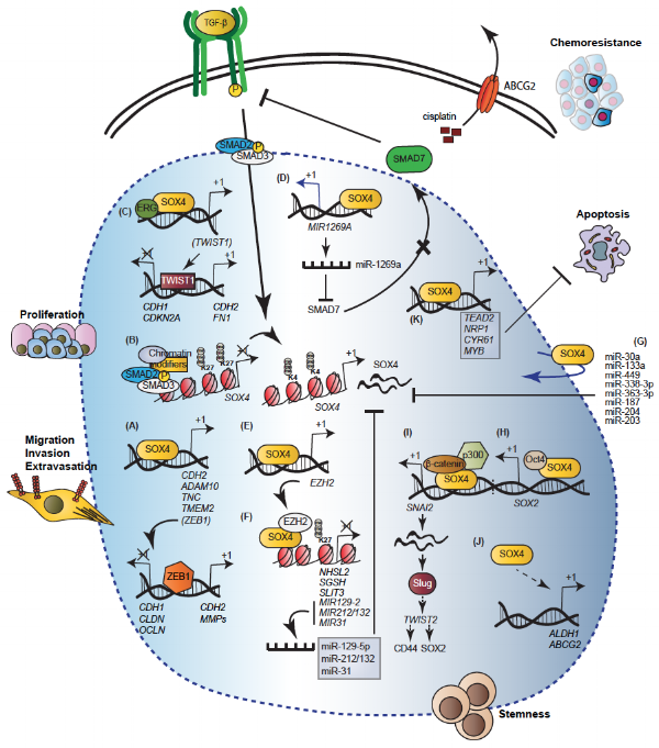
SOX4: Joining the Master Regulators of Epithelial-to-Mesenchymal Transition?

SOX4 EMT

The epithelial-to-mesenchymal transition (EMT) is an important developmental program exploited by cancer cells to gain mesenchymal features. Transcription factors globally regulating processes during EMT are often referred as 'master regulators' of EMT, and include members of the Snail and ZEB transcription factor families. The SRY-related HMG box (SOX)4 transcription factor can promote tumorigenesis by endowing cells with migratory and invasive properties, stemness, and resistance to apoptosis, thereby regulating key aspects of the EMT program. In this review recently published in Trends in Cancer, we argue that SOX4 should also be considered as a master regulator of EMT, and review the molecular mechanisms underlying its function.

Congratulations Dr Luca Braccioli - PhD Survivor !

Luca Braccioli.jpg

Transcriptional regulation and cellular strategies in neuroregeneration

Neurogenesis and gliogenesis are processes that occur during development of the CNS as well as after insult of the nervous system. These tightly regulated processes take place in specialized niches during embryogenesis and adulthood to generate functional cells, starting from defined progenitor cells such as neural stem cells (NSCs). During his PhD, Luca took a multifaceted experimental approach to better understand NSC biology both in vitro and in vivo. Transcriptional regulation is essential since the expression of specific genes is the key to control timing and fate of the differentiation process.

How do NSC work? In Luca Braccioli's thesis two NSC transcriptional regulators, Foxp1 and Sox4 are shown to be crucial for control of neurogenesis and gliogenesis respectively. Mutations in the Foxp1 gene have been associated with speech defects, autism and other intellectual disabilities, as well as being defined as necessary for neurogenesis. In the work presented in this thesis, Luca sought to define the molecular mechanisms mediated by Foxp1 that regulate NSC differentiation. Sox4 has been described as inhibitor of gliogenesis and myelination in oligodendrocyte precursor cells. In this context we investigated the role and molecular mechanisms underlying Sox4-mediated regulation of oligodendrogenesis.

Repairing brain damage. The use of cellular strategies to repair CNS insults has been widely investigated in recent years. During his PhD Luca further evaluated the therapeutic potential of NSCs and MSCs to treat perinatal hypoxic-ischemic brain damage (HI). Moreover, he examined the hypothesis of the use of exosomes as a cell-free alternative as a therapeutic option upon brain insults and HI.

A tumor suppressor role for C/EBPα in solid tumors: more than fat and blood?

C/EBPalpha

The transcription factor CCAAT/enhancer-binding protein alpha (C/EBPα) plays a critical role during embryogenesis and is thereafter required for homeostatic glucose metabolism, adipogenesis and myeloid development. Its ability to regulate the expression of lineage-specific genes and induce growth arrest contributes to the terminal differentiation of several cell types, including hepatocytes, adipocytes and granulocytes. CEBPA loss of-function mutations contribute to the development of ~10% of acute myeloid leukemia (AML), stablishing a tumor suppressor role for C/EBPα. Deregulation of C/EBPα expression has also been reported in a variety of additional human neoplasias, including liver, breast and lung cancer. However, functional CEBPA mutations have not been found in solid tumors, suggesting that abrogation of C/EBPα function in non-hematopoietic tissues is regulated by alternative mechanisms. Here, we discuss the function of C/EBPα in solid tumors, focussing on the molecular mechanisms underlying its tumor suppressive role. This review was recently published in Oncogene

ANKZF1: a novel gene associated with infantile-onset inflammatory bowel disease

ANKZF1.png

Infantile-onset inflammatory bowel disease (IO IBD) is an invalidating illness with an onset before 2 years of age and has a complex pathophysiology in which genetic factors are important. Here using homozygosity mapping and whole-exome sequencing, Desiree Haaften-Visser identifies for the first time mutations in the ANKZF1 gene in IO IBD patients. Although the function of ANKZF1 in mammals had not been previously evaluated, ANKZF1 was found to have an indispensable role in the mitochondrial response to cellular stress. ANKZF1 is located diffusely in the cytoplasm and translocates to the mitochondria upon cellular stress. ANKZF1 depletion reduces mitochondrial integrity and mitochondrial respiration under conditions of cellular stress. The ANKZF1 mutations identified in IO IBD patients results in dysfunctional ANKZF1, as shown by an increased level of apoptosis in patients' lymphocytes, a decrease in mitochondrial respiration in patient fibroblasts with a homozygous ANKZF1 R585Q mutation, and an inability of ANKZF1 R585Q and E152K to rescue the phenotype of yeast deficient in Vms1, the yeast homologue of ANKZF1. These data indicate that loss-of-function mutations in ANKZF1 result in deregulation of mitochondrial integrity, and this may play a pathogenic role in the development of IO IBD. This work has been recently published in the Journal of Biological Chemistry.